Elbow Dysplasia
in Dogs
Fixed prices. Fixed pets. 
What is canine elbow dysplasia?
Elbow dysplasia is a term for a group of conditions which affect the elbow joint when the dog is growing.
The elbow joint is formed by the humerus, radius and ulna bones. The conditions included under elbow dysplasia are ununited anconeal process (UAP), fragmented medial coronoid process (FMCP), osteochondritis dissecans (OCD) and joint incongruity.
These can be seen in isolation or in combination. All of these conditions will lead to secondary osteoarthritis developing in the joint.
Both the primary condition and the secondary osteoarthritis can cause lameness, so early investigation and treatment can be beneficial.

What are the signs and symptoms
of elbow dysplasia?
Elbow dysplasia is first seen when the dog is growing and signs can be seen from about four months of age. Adults dogs can also present with the disease due to the secondary osteoarthritis that develops in the joint.
Medium to large breeds are most commonly affected. Breeds with a high incidence of elbow dysplasia include Labrador retrievers, Golden retrievers, Rottweilers, German shepherd dogs, Bernese Mountain Dogs and Dogue de Bordeaux.
The most common sign of elbow dysplasia is forelimb lameness. This is often worse when the dog gets up from rest or after heavier exercise.
How is it diagnosed?
The diagnosis of elbow dysplasia is based on a combination of findings on examination and imaging.
The signalment – breed, age, sex – and history are also useful.
Clinical Examination
Examination findings include forelimb lameness, pain on manipulation of the elbow joint and associated fluid swelling.
The affected elbow can become thickened and develop crepitus (crunchiness on manipulation) due to osteoarthritis. The disease can be bilateral (affecting both legs) in some patients.
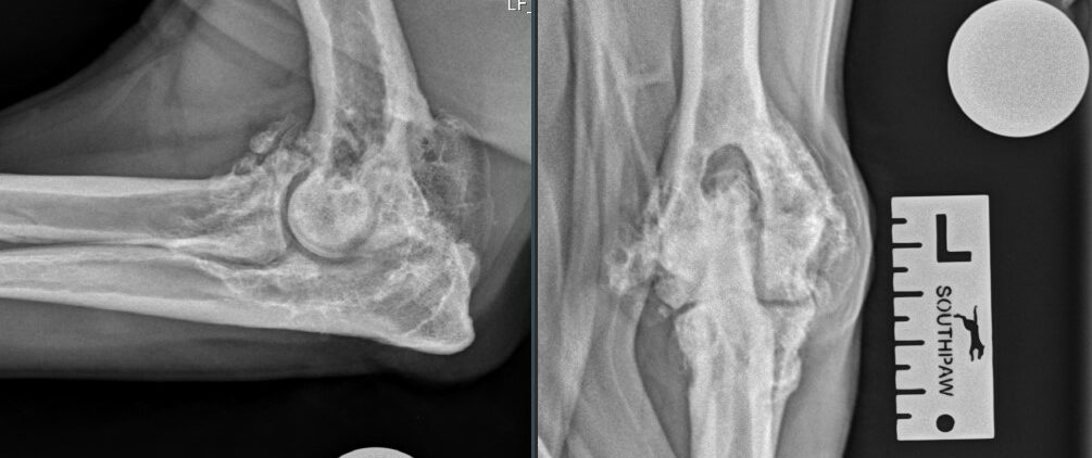
Radiography
Radiography or x-ray is of varying benefit in the diagnosis of elbow dysplasia. Some of the underlying conditions can be easily identified such as ununited anconeal process.
Other components, such as fragmented medial coronoid process and incongruity, are harder to identify due to the bones of the joint overlying each other.
In these cases advanced imaging is required to confirm the diagnosis. Radiography is good for identifying the secondary osteoarthritis.
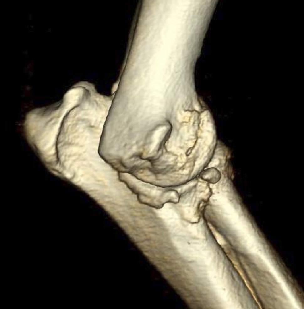
CT Scan
CT (computed tomography) gives much more detailed images of the elbow joint and overcomes the problem of the overlying bones.
Three-dimensional reconstructions of the bones can also be obtained. CT is the best modality for assessing the medial coronoid process and incongruity.
Arthroscopy
Arthroscopy is the direct visualisation of the inside of a joint using a small rigid camera, only a couple of millimetres in diameter.
This allows examination of the medial coronoid process for fragmentation, the humeral condyle for OCD and overall assessment of the cartilage. It also allows for therapeutic management of some of these conditions.
Treatment options?
There are a range of possible treatments for elbow dysplasia depending on the underlying condition,
age at presentation and degree of osteoarthritis.
Conservative
This includes the following list, but is not exhaustive, and the best choice for your pet will be discussed with you following the investigation.
- Exercise management
- Weight control
- Physiotherapy
- Hydrotherapy
- Nutraceuticals (glucosamine)
- Non steroidal anti-inflammatories
- Intra-articular medications – platelet therapy,
- stem cell therapy, steroids
Surgical
Some of the primary conditions can be treated surgically and the type of surgery is dependent on which diagnosis has been made. There are also surgical options for managing the osteoarthritic change if this is not responsive to conservative management.
- Fixation of an ununited anconeal process with a screw
- Debridement (removal) of the cartilage flap from an OCD lesion – arthroscopically or open surgery
- Removal of a fragmented coronoid process -arthroscopically or open surgery
- Osteotomy (bone cutting) of the ulna to remove pressure from certain areas of the elbow joint
- Osteotomy and then special plating of the ulna to shift how weight is transferred across the elbow – PAUL surgery
- Partial resurfacing of the elbow joint – CUE surgery
- Total elbow replacement
Frequently Asked Questions
What are the causes of elbow dysplasia?
Elbow dysplasia is a multifactorial disease and there is a genetic predisposition in affected dogs. Multiple genes are involved and there is not direct transmission, so not all dogs that are genetically affected will develop disease.
Joint congruity is the most important factor in determining which form of elbow dysplasia develops. Exercise level and diet can influence the severity of disease but do not cause it.
Can elbow dysplasia be fixed?
Unfortunately elbow dysplasia can not be cured, but in most cases the disease can be managed to achieve a quality of life that is acceptable for you and your pet.
What can help elbow dysplasia?
There are a range of possible managements depending on the primary cause and level of secondary osteoarthritis. The options will be discussed with you following the investigation stage to decide on the best treatment plan for your pet.
Is elbow dysplasia painful?
Both the primary conditions and osteoarthritis can cause inflammation in the affected elbow joint which will be manifested as lameness.
How can I exercise my dog with elbow dysplasia?
The level of exercise your pet can do will depend on the stage of disease, the degree of osteoarthritis and whether any specific surgical procedures have been carried out.
The aim of any management, conservative or surgical, is to get you pet back to best level of activity possible, but unfortunately some dogs may have a degree of persistent lameness and their exercise will need to be managed accordingly.
How successful is surgery for elbow dysplasia?
The outcomes from any surgery and the risks of complication are dependent on the type of surgery that is carried out and this will be discussed with you before any procedure is undertaken.
How long is the recovery for elbow dysplasia surgery in dogs?
The recovery period from any surgery will depend on the type of surgery that is carried out and the post-operative regime will be discussed with you prior to any surgery. Some procedures will require longer periods of rest and confinement than others.
Customer Reviews
Arranging a referral for your pet?
If your dog has been diagnosed with elbow dysplasia or has a forelimb lameness and you would like us to carry out further investigations and treatment all you have to do is ask your veterinary surgeon to refer you to us.
They can do this easily via our website, by email or by phone call. If you can let us know when you have made the request we will know to expect the referral and can follow it up if required.
The Old Dairy Yard, Welsh Way, Ready Token, Cirencester, Gloucestershire GL7 5SY